BIO

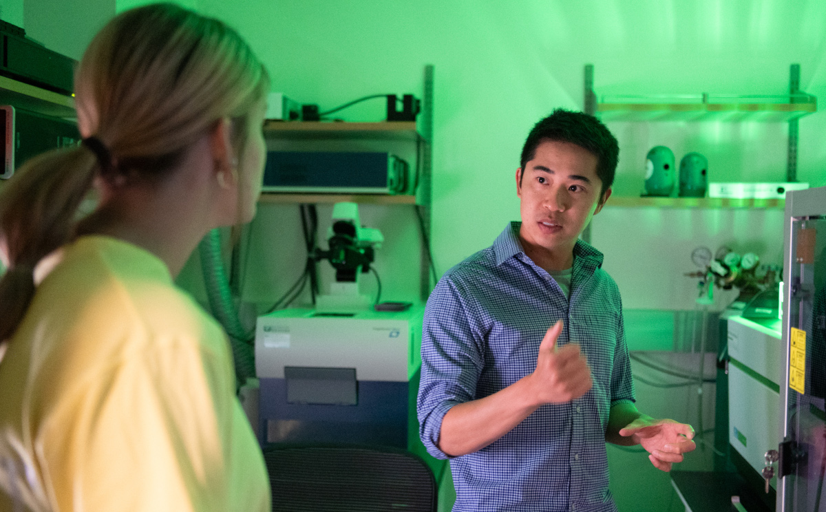
I am a Core Bioinformatics Scientist II at the Van Andel Institute, where I build software to extract meaningful data from complex biological images. Within the Optical Imaging Core, my work focuses on developing tools to analyze and visualize massive, multi-dimensional microscopy datasets.
My research sits at the intersection of biology and computation. I utilize advanced microscopy to capture the dynamics of living cells over extended periods, while simultaneously building the software needed to process these phenomena. Together with VAI collaborators, I apply these methods to better understand the mechanisms of human diseases, with a primary focus on cancer and Parkinson’s disease. I am currently focused on the frontier of image informatics, specifically leveraging AI and machine learning to automate and enhance data extraction and understanding.
Previously, I served as the Biological Image Analysis Group Leader at the University of Colorado Boulder. There, I led the Bioimaging Lab — a collaborative undergraduate research group — and taught Quantitative Optical Imaging across the Departments of Biochemistry and Molecular, Cellular, and Developmental Biology.
EDUCATION & EXPERIENCE
-
Van Andel Institute
Optical Imaging Core
2025 - PresentCore Bioinformatics Scientist II
-
University of Colorado Boulder
BioFrontiers Institute
2024 - 2025Biological Image Analysis Group Leader
- Co-lecturer: Quantitative Optical Imaging
- PI: Bioimaging Lab
-
University of Colorado Boulder
BioFrontiers Institute
2016 - 2024Image Analysis Specialist
-
University of Colorado
Department of Electrical Engineering
2014 - 2016Postdoctoral Research Associate
-
Washington University in St Louis
Department of Biomedical Engineering
2011 - 2014Postdoctoral Research Associate
- Photoacoustic wavefront shaping
-
University of Otago
Department of Physics
2007 - 2011PhD
- Optical detection of ultrasound
- Spatial homodyne detection
-
University of Otago
Department of Physics
2003 - 2006BSc (Hons I)
- Ion trapping in high-finesse optical traps
RECENT PUBLICATIONS
-
Calcite precipitation by the nitrogen-fixing cyanobacterium Anabaena sp. ATCC 33047
Christian M. Brininger, Jian Wei Tay, Evan B Johnson, Erin Espelie, and Jeffrey C. Cameron.
Communications Biology • 2025 -
Machine learning models for segmentation and classification of cyanobacterial cells
Clair A. Huffine, Zachary L. Maas, Anton Avramov, Christian M. Brininger, Jeffrey C. Cameron, and Jian Wei Tay.
Photosynthesis Research • 2025 -
The Spatial Distribution of Lipophilic Cations in Gradient Copolymers Regulates Polymer–pDNA Complexation, Polyplex Aggregation, and Intracellular pDNA Delivery
Jessica L. Lawson, Ram Prasad Sekar, Aryelle R. E. Wright, Grant Wheeler, Jillian Yanes, Jordan Estridge, Chelsea G. Johansen, Nikki L. Farnsworth, Praveen Kumar, Jian Wei Tay, and Ramya Kumar.
Biomacromolecules • 2024